
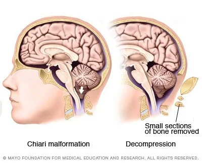
Chiari Malformation is crowding of the lower portion of the brain in the base of the skull.
how common
There are more than 200,000 cases per year in the US
surgeries
Dr. Thompson has operated on over 230 Chiari patients.
complcations
―
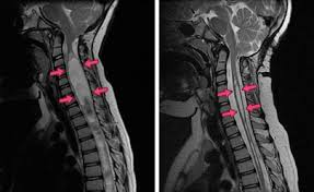
Crowding of the brain in Chiari Malformation leads to the downward protrusion of the cerebellar tonsils through the foramen magnum (the opening of the skull base) which interrupts normal cerebral spinal fluid flow. In 25% of patients a Syringomyelia is present. Syringomyelia is a chronic progressive degenerative disorder characterized by a fluid-filled cyst located in the spinal cord. It is usually seen in the cervical region but can extend downward into the thoracic or lumbar segments. The exact development of syringomyelia is unknown but many theories suggest that the herniated tonsils in Chiari malformation (type I) form a "plug" which does not allow an outlet of CSF from the brain to the spinal canal.
There are 2 classifications of Chiari Malformations
Type I: small posterior fossa due to a multitude of causes.
Type 2: Associated with myelomeningocele and multiple brain anomalies, hydrocephalus and syringomyelia are both very common.
Both types present with similar symptoms. Symptoms vary greatly
Headaches, usually located in the back of the head especially with coughing. headaches may be associated with nausea or vomiting.
Weakness, numbness, or tingling of extremities and/or hyperactive reflexes
Unsteadiness, gait or coordination disturbance
Inability to feel changes in the temperature
Cranial nerve palsies or changes in vision
Sexual dysfunction, and loss of bowel and bladder control
Plan of care:
workup
Diagnosis is typically made through a combination of patient history and neuroimaging, such as an MRI.
treatment
Surgical treatment options
“Bony Only” Chiari Decompression with cervical laminectomy of C1
An incision is made along the base of the skull, and a window of bone at the base of the skull is removed. The “roof” of the first cervical vertebrae is also removed. There is also a small ligament that is removed to ensure there is no band-like tension at the base of the skull. Typical hospital stay: ICU 1 night, Floor-1 to 3 nights
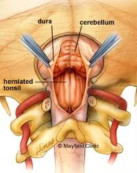
Chiari Decompression with Duraplasty
After the bony decompression is completed, the covering of the brain (DURA) is opened under a microscope. The cerebellar tonsils are in view, and an electro-cautery tool is used to shrink this tissue. This is a safer method than cutting the tissue out as it minimizes bleeding risk. Think shrink wrap... The tonsils are like the “appendix of the brain” in that they serve no neurological function. The tissue has been squeezed for so long as well, that it is causing more harm than good. Micro-instruments are then used to cut away and intradural adhesions or scarring within the arachnoid layer. We then harvest an autograft, using periosteum from the patient’s own skull. This is then sutured in a water-tight fashion under the microscope. We then place some fibrin glue over this to ensure there is no leaking of spinal fluid. The fascia is then closed in a watertight fashion as well. Typical hospital stay: ICU 1 night, stepdown 1 night, Floor-1 to 2 nights.
follow up
Typically will see patient at 2 weeks, 6 weeks, and 3 months post-op. Will typically get a new MRI scan at the 3 month visit to re-evaluate. Children are usually yearly for 1-2 years then seen in the office as needed. Adults are seen as needed unless they had a syrinx, in which case yearly imaging would be needed to ensure resolution of the syrinx. The syrinx is not surgically corrected as fixing the main source (the crowding at the base of the skull) allows for normal CSF flow through the central canal and resolution of the syrinx naturally.
Want to connect with other local Chiari patients? Click below to join our private Facebook group.