
Hydrocephalus is the buildup of too much cerebrospinal fluid in the brain. When you have too much, it puts harmful pressure on your brain.
Site
68 Holloway Rd.
Installed
Dr. Thompson has performed over 600 shunt operations.
Team
Cherise King
Justin Gomez
―
Hydrocephalus develops when excess cerebrospinal fluid (CSF) builds up within the ventricles (fluid-containing cavities) of the brain. When the ventricles swell, they cause harmful pressure on the tissues of the brain.
Hydrocephalus can be congenital, or present at birth. Causes include genetic problems and problems with how the fetus develops. An unusually large head is the main sign of congenital hydrocephalus.
Hydrocephalus can also happen after birth. This is called acquired hydrocephalus. It can occur at any age. Causes can include head injuries, strokes, infections, tumors, and bleeding in the brain.
2. Arachnoid Cyst: the arachnoid is one of the coverings of the brain and contains CSF, small pockets of fluid can become encapsulated and does not communicate with the remaining CSF
Symptoms in adults and children include:
- Headaches
- Nausea and vomiting
- Drowsiness, lethargy
- Gait or balance problems
- Blurry vision
- Thinking and memory problems
Babies/Young children are often unable to verbalize symptoms therefore it is important to report any of the following concerns:
- Bulging fontanel
- Increased head circumference
- Irritability, lethargy
- Unexplained vomiting
- Developmental delay
Hydrocephalus can permanently damage the brain, causing problems with physical and mental development. If untreated, it is usually fatal. With treatment, many people lead normal lives with few limitations. Treatment usually involves surgery to insert a shunt. A shunt is a flexible but sturdy plastic tube. The shunt moves the cerebrospinal fluid to another area of the body where it can be absorbed.
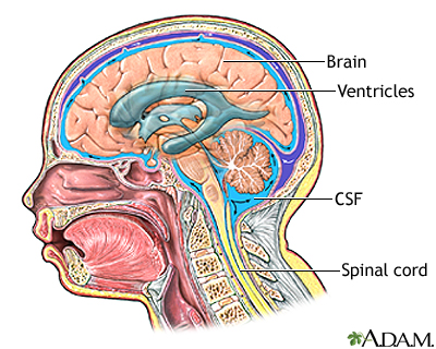
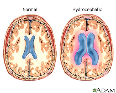
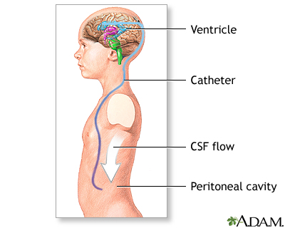

Ventriculoperitoneal shunting is a procedure performed in the operating room under general anesthesia. A flap is cut in the scalp and a small hole is drilled in the skull. A small catheter is passed into a ventricle of the brain. A pump is attached to the catheter to keep the fluid away from the brain. Another catheter is attached to the pump and tunneled under the skin, behind the ear, down the neck and chest and into the peritoneal cavity (abdominal cavity). The CSF is absorbed in the peritoneal cavity.
Plan of Care:
1. Work-up
Diagnosis of hydrocephalus begins with imaging. In neonates a head ultrasound is used to assess the size of the ventricles. For babies and children workup includes a CT scan/MRI of the head. Dr. Thompson prefers a fast-spin MRI to avoid radiation exposure and possible complications associated with anesthesia for a sedation MRI.
2. Treatment
Ventriculoperitoneal shunting is often crucial for preventing serious brain damage in children with hydrocephalus. Common problems associated with VP shunts include shunt malfunction and shunt infection. Shunts are generally left in place for many years if they function without problems.
Neuroendoscopic third ventriculostomy-a very small camera is inserted into the ventricles and a small hole is made in the bottom of the ventricle, creating a new pathway for CSF diversion.
Neuroendoscopic fenestration of an arachnoid cyst- a very small camera is inserted into cyst and opened to communicate with the remaining CSF
3. Follow-up
Typical follow up includes imaging to assess the size of the ventricles.